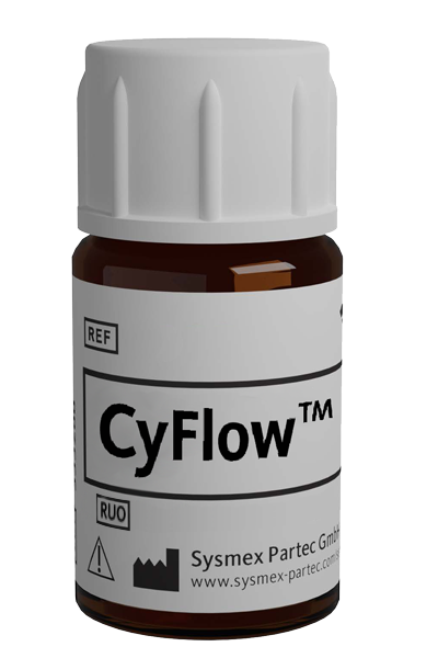
CyFlow™ CD3 PE

| Alternative Name: | Leu4, T3 |
| Antibody: | Yes |
| Antigen: | CD3 |
| Application: | Flow cytometry |
| Clonality: | monoclonal |
| Clone: | MEM-57 |
| Emission Maximum: | 576 nm |
| Excitation Maximum: | 496 nm, 565 nm |
| Field of Interest: | Immunophenotyping |
| Format/Fluorochrome: | PE |
| Isotype: | IgG2a |
| Laser: | Blue , Green, Yellow |
| Regulatory Status: | RUO |
| Source Species: | Mouse |
| Target Species: | Human |
| Product number: | BY096263 |
For Research Use Only
| HLDA Workshop | HLDA IV—WS Code T 96 |
| Quantity | 100 tests |
| Volume | 2.0 mL |
| Immunogen | Human thymocytes and T lymphocytes |
| Background Information | CD3 complex is crucial in transducing antigen-recognition signals into the cytoplasm of T cells and in regulating the cell surface expression of the TCR complex. T cell activation through the antigen receptor (TCR) involves the cytoplasmic tails of the CD3 subunits CD3γ, CD3 δ, CD3ε and CD3ζ. These CD3 subunits are structurally related members of the immunoglobulins superfamily encoded by closely linked genes on human chromosome 11. The CD3 components have long cytoplasmic tails that associate with cytoplasmic signal transduction molecules. This association is mediated at least in part by a double tyrosine-based motif present in a single copy in the CD3 subunits. CD3 may play a role in TCR-induced growth arrest, cell survival and proliferation. The CD3 antigen is present on 68-82% of normal peripheral blood lymphocytes, 65-85% of thymocytes and Purkinje cells in the cerebellum. It is never expressed on B or NK cells. Decreased percentages of T lymphocytes may be observed in some autoimmune diseases. |
| Usage | The reagent is designed for Flow Cytometry analysis of human blood cells. Recommended usage is 20·µl reagent·/ 100·µl of whole blood or 10^6 cells in a suspension. The content of a vial (2 ml) is sufficient for 100 tests. |
| Storage Buffer | The reagent is provided in stabilizing phosphate buffered saline (PBS) solution, pH ≈7.4, containing 0.09% (w/v) sodium azide. |
| Storage | Avoid prolonged exposure to light. Store in the dark at 2-8°C. Do not freeze. |
| Stability | Do not use after expiration date stamped on vial label. |
| McMichael AJ, Beverley PCL, Cobbold S, et al (Eds): Leucocyte Typing III, White Cell Differentiation Antigens. Oxford University Press, Oxford. 1987; 1‑1050. < NLM ID: 8913266 > | Horejsí V, Angelisová P, Bazil V, Kristofová H, Stoyanov S, Stefanová I, Hausner P, Vosecký M, Hilgert I: Monoclonal antibodies against human leucocyte antigens (II); Antibodies against CD45 (T200), CD3 (T3), CD43, CD10 (CALLA), transferrin receptor (T9), a novel broadly expressed 18‑kDa antigen (MEM‑43) and a novel antigen of restricted expression (MEM‑74). Folia Biol (Praha). 1988; 34(1):23‑34. < PMID: 2968928 > | Knapp W, Dorken B, Gilks W, Rieber EP, Schmidt RE, Stein H, von dem Borne AEGK (Eds): Leucocyte Typing IV. Oxford University Press, Oxford. 1989; 1‑1820. < NLM ID: 8914679 > | Soucek J, Chudomel V, Hrubá A, Lindnerová G: Induction of NK and LAK activities in human lymphocyte culture by a cytosol fraction from leukemic myeloblasts and by monoclonal antibody CD 3. Neoplasma. 1991; 38(1):33‑41. < PMID: 2011208 > | Hilgert I, Franĕk F, Stefanová I, Kaslík J, Jirka J, Kristofová H, Rossmann P, Soucek J, Horejsi V: Therapeutic in vivo use of the A1‑CD3 monoclonal antibody. Transplantation. 1993; 55:435. < PMID: 8434399 > | Soucek J, Hilgert I, Budová I, Lindnerová G: Augmentation of NK cell activity and proliferation in cultured lymphocytes of leukemic patients by monoclonal antibodies CD3 and interleukin‑2. Neoplasma. 1994; 41(2):75‑81. < PMID: 8208318 > | Dave VP, Cao Z, Browne C, Alarcon B, Fernandez-Miguel G, Lafaille J, de la Hera A, Tonegawa S, Kappes DJ: CD3 delta deficiency arrests development of the alpha beta but not the gamma delta T cell lineage. EMBO J. 1997 Mar 17; 16(6):1360‑70. < PMID: 9135151 > | Panyi G, Bagdány M, Bodnár A, Vámosi G, Szentesi G, Jenei A, Mátyus L, Varga S, Waldmann TA, Gáspar R, Damjanovich S: Colocalization and nonrandom distribution of Kv1.3 potassium channels and CD3 molecules in the plasma membrane of human T lymphocytes. Proc Natl Acad Sci USA. 2003 Mar 4; 100(5):2592‑7. < PMID: 12604782 > | Brdicková N, Brdicka T, Angelisová P, Horváth O, Spicka J, Hilgert I, Paces J, Simeoni L, Kliche S, Merten C, Schraven B, Horejsí V: LIME: a new membrane Raft‑associated adaptor protein involved in CD4 and CD8 coreceptor signaling. J Exp Med. 2003 Nov 17; 198(10):1453‑62. < PMID: 14610046 > | Huang Y, Wange RL: T cell receptor signaling: beyond complex complexes. J Biol Chem. 2004 Jul 9; 279(28):28827‑3. < PMID: 15084594 > | Kuhns MS, Davis MM, Garcia KC: Deconstructing the form and function of the TCR/CD3 complex. Immunity. 2006 Feb; 24(2):133‑9. < PMID: 16473826 > | Alarcón B, Swamy M, van Santen HM, Schamel WW: T‑cell antigen‑receptor stoichiometry: pre‑clustering for sensitivity. EMBO Rep. 2006 May; 7(5):490‑5. < PMID: 16670682 > | Drbal K, Moertelmaier M, Holzhauser C, Muhammad A, Fuertbauer E, Howorka S, Hinterberger M, Stockinger H, Schütz GJ: Single‑molecule microscopy reveals heterogeneous dynamics of lipid raft components upon TCR engagement. Int Immunol. 2007 May; 19(5):675‑84. < PMID: 17446208 > | Kanderova V, Kuzilkova D, Stuchly J, Vaskova M, Brdicka T, Fiser K, Hrusak O, Lund‐Johansen F, Kalina T: High‐resolution antibody array analysis of childhood acute leukemia cells. Mol Cell Proteomics. 2016 Apr 1; 15(4):1246‐61. < PMID: 26785729 >
